B.Sc ZOOLOGY Topic :- HERDAMINA (Paper :- Chordate Structure and Function)
HERDMANIA
CLASSIFICATION -
Kingdom - Animalia
Phylum - Chordat
Class - Ascidiacea
Order - Stolidobranchi
Family - Pyurude
Genus - Herdmani
Subphylum Urochordata (Gr., uros = tail; chorde = cord) includes a peculiar group of widely distributed marine animals called sea squirts or ascidians and their allies. In most of them several chordate characters are lost in the adults, but their chordate affinities are clearly seen in their free-swimming larvae which have pharyngeal gill-clefts, a dorsal tubular central nervous system, and a notochord which is confined only to the tail (hence, the name Urochordata).
During metamorphosis the chordate characters are lost except gill-clefts. Gill-clefts do not open to the exterior but into an ectoderm-lined atrium. The body becomes surrounded by a coat called test or tunic (hence, the name Tunicata). The changes in metamorphosis are a specialisation due to a sedentary life in which ciliary feeding is perfected, due to which locomotory organs and neuro-sensory system are simplified and reduced.
They have no metameric segmentation and a coelom is absent. They are also hermaphrodite. About 2,200 Urochordata are known, they show a great diversity of structure, habits and habitats. They may be sessile or free-swimming. They are found in all seas, being found along the shores and up to a depth of more than 3 kms.
The genus Rhabdocynthia was first established by Herdman in 1891. But, in 1910, Hartmeyer changed it to Herdmania according to the law of priority as it was originally proposed by Lahille in 1888.
Habits and Habitat:-

Herdmania pallida is a solitary marine form found in shallow waters along the Indian sea coast. Each animal is found attached to the substratum usually separately, at its postero-ventral end by means of a foot. When the substratum is sandy, an expanded or elongated foot is formed which remains imbedded in the sand and keeps the animal fixed to the sea bottom. A large number of organisms inhabit the test of Herdmania, some of these are merely attached to its surface, while others are more or less imbedded within its substance.
Sometime an individual is found attached to a living gastropod shell (e.g., Turbinella pyrum, T. rapa, Xancus, etc.) showing commensalism. Herdmania protects the gastropod from enemies since it is unpalatable due to its spicules, and gastropod takes it from place to place so that it may get good food and oxygen, etc. The food of Herdmania consists of microscopic plants and animals, e.g., diatoms, algae and infusorians.
Herdmania is more or less widely distributed, though it cannot be called a cosmopolitan species. H. pallida having been recorded from the Indian, the Pacific and the Atlantic Oceans, as also from Malaya and the West Indies. Herdmania is hermaphrodite and oviparous. Fertilisation takes place in the sea and the tadpole larvae emerging from the eggs lead a short free-swimming life before they settle down to the sea-bottom to lead a fixed existence after going retrogressive metamorphosis.
EXTERNAL MORPHOLOGY :-
1.Shape, Size and Colouration:-
The body of Herdmania pallida is roughly oblong in outline, narrower at its attached than at its free end. At its free end, it is provided with two external openings—the branchial and atrial apertures. The average size of the adult is about 9.5 cm long, 7 cm broad and 4 cm thick. Older animals may even attain to a size of 12 x 8 x 4 cm, while an exceptionally large measures 13 x 8 x 4.5 cm. The foot, when present, may attain a length of as much as 3 to 4 cms. The general colour of the body in a fresh specimen is pink.
The presence of bright red patches, formed of terminal knobs (ampullae) in the blood vessels of the test, is a characteristic feature of Herdmania. The test is soft and leathery. It is more or less transparent in a young animal, but in an adult becomes usually opaque. The general surface of the test is much corrugated all over with lines, some shallow and others fairly deep, running in a criss-cross manner.

2. Body Divisions:-
It is the distal free portion of the body. The branchial aperture marks the anterior end of the animal; consequently the opposite end attached to the substratum is the posterior end. The side on which the atrial aperture is placed marks the dorsal side of the body, which is very limited. The side opposite to that on which the atrial opening is placed and which is partly attached to the substratum is ventral side which is extensive.
The branchial and atrial apertures are situated on short protuberances of the body called branchial and atrial siphons respectively. When fully extended, the atrial siphon is longer than the branchial. In a large-sized animal the atrial siphon measures about 1.5 cm, while the branchial siphon is only about 1 cm in length.
The atrial siphon is almost always directed upwards, the atrial aperture being more or less upright, while the branchial siphon is always bent a little outwards and the branchial aperture opens more or less laterally, always directed away from the atrial aperture. The average diameter of the branchial aperture, when fully expanded, is about 2 cm and that of the atrial aperture about 1.2 cm.
At the base of branchial siphon, there is a ring of long branchial tentacles, while at the base of the atrial siphon there is a ring of slightly serrated folds which constitute the atrial tentacles. The test of the siphons is very elastic and can contract to close the apertures at the slightest disturbance of sea water.
2.Foot:-
The foot, when present varies in character according to the nature of the substratum which the animal inhabits. If the substratum is of fine sand, the foot has an oval shape and a smooth surface and the test is quite hard in consistency. But if the substratum consists of coarse and the broken shell-pieces, the foot is irregular in outline and more or less soft in consistency.
3.Test or Tunic:
The external covering surrounding the animal is a leathery, translucent test or tunic composed of tunicin, a substance akin to cellulose of plants. The foot is made entirely of test. It acts as a receptor as well as a respiratory organ. It is about 4-8 mm thick. It is ectodermal in origin.
The test has:
(1) A clear matrix in which is embedded,
(2) Cells of various shapes,
(3) Interlacing fibrils,
(4) Calcareous spicules, and
(5) Branching vascular vessels.
(1) Matrix:-It is gelatinous and made of a polysaccharide called tunicine.
(2) Cells:-These are mesodermal in origin and have migrated into the test.
These are of 6 or 7 different types:
(a) Large eosinophilous cells,
(b) Small eosinophilous cells,
(c) Small amoeboid cells,
(d) Granular cells,
(e) Round vacuolated cells,
(f) Nerve cells with several processes, and
(g) Squamous epithelial cells.
(3) Interlacing fibrils:
These form a fine network in the test and resemble with smooth muscle fibres.
(4) Calcareous spicules:
Herdmania has large number of calcareous spicules of two types. All the spicules bear several equidistant rings of minute spines all pointing in the same direction all along their length
(5) Blood vessels:
These blood vessels form a network in the test and end in bulb-like dilations called vascular ampullae. The vascular ampullae form vascular areas of bright red patches on the test. Vascular vessels and ampullae transport blood and, thus bring food to the test; they also act as accessory respiratory organs. The ampullae are also receptor organs.
4.Mantle or Body Wall:-
Inside the test is the body wall or mantle. The mantle secretes the test. The mantle is present beneath the test and is attached only at the branchial and atrial apertures where it forms branchial and atrial siphons.
The mantle encloses a large atrial cavity or Prebranchial zone atrium, containing water. Histologically the mantle has an outer layer of ectoderm, middle layer of mesoderm, and an inner layer of endoderm forming the outer lining of atrial cavity.
1.. Outer Ectoderm:
It is formed of a single layer of flat, hexagonal cells. It turns inside at pericardium the branchial and atrial apertures and extends up to the base of the siphons forming stomodaeum and proctodaeum respectively.

2. Inner Ectoderm:
It is formed of single layer of flat polygonal cells and forms the lining of the atrium.
3. Middle Mesoderm:
It lies beneath the outer ectoderm. It is composed of connective tissue traversed by muscle fibres, blood sinuses and nerve fibres.
The muscle fibres are of unstriped type and are arranged in three sets:
(i) Around the branchial and atrial siphons the mantle has annular muscles in several rings.
(ii) Below the annular muscles are longitudinal muscles which start from branchial and atrial apertures, then fan out up to the middle of the body on each side.
(iii) Branchioatrial muscles found in between two siphons.
Digestive System:-
Alimentary Canal:-
The alimentary canal is coiled, beginning from the mouth and ending in anus. It has the following parts:-
1. Mouth:-
The mouth or branchial aperture lies at the top of branchial siphon, which is designated as the anterior end of the body. It is guarded by four lips (lobes) of the test. The mouth leads into a branchial siphon.
2. Branchial Siphon:-

It is a narrow and tubular cavity lined by ectoderm and is called the stomodaeum or buccal cavity. At the base of the stomodaeum is a ring of branching tentacles which act as a sieve allowing only minute food particles to go in. These are richly innervated and their number is about 64.
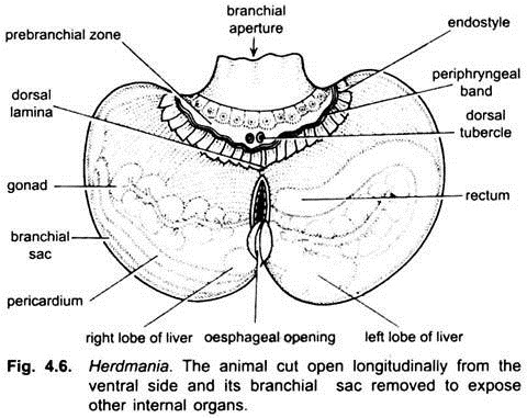
Below the tentacles is a smooth prebranchial (prepharyngeal) zone having a swollen dorsal tubercle made of two spiral coils. Then there are two thin ciliated ridges, the peripharyngeal bands lying parallel and encircling the upper end of the pharynx. The posterior one of the two peripharyngeal bands is joined mid-dorsally to a dorsal lamina, and mid-ventrally with an endostyle.
3. Pharynx:
It occupies the major part of the body cavity and is differentiated into two- prebranchial zone and branchial sac.
(i) Prebranchial Zone:
It is the smaller anterior region having smooth walls without folds, cilia and stigmata or gill-slits.
ADVERTISEMENTS:
(ii) Branchial Sac or Pharynx:
It is the posterior larger part of the pharynx. The pharynx almost fills the atrial cavity. The wall of the pharynx is perforated by several rows of stigmata arranged in transverse rows, through these the cavity of the pharynx communicates with the atrial cavity. The edges of stigmata are beset with cilia which drive currents of water from the pharynx into the atrial cavity.
The stigmata are not gill-clefts, but they are formed by sub-divisions of a few original gill-clefts. Between the stigmata the wall of the pharynx has transverse and longitudinal bars containing blood vessels, thus, the pharynx appears like a basket. The lining of the pharynx is raised into ten longitudinal folds on each side.
(a) Dorsal Lamina:
Dorsal lamina is a thin flap lying inside the mid-dorsal roof of the pharynx. It bears a number of conical, ciliated projections called languets. The dorsal lamina runs from the posterior peripharyngal band to the opening of the oesophagus. The dorsal lamina and its languets can bend to one side to form a sort of tube through which food-laden mucus passes.
(b) Endostyle:
Lying in the mid- ventral floor of the pharynx is an endostyle. It is a longitudinal groove with four longitudinal rows of gland cells with ciliated cells between them. The middle row of cells has long cilia. The gland cells of endostyle secrete mucus which is driven laterally by ciliated cells. Endostyle is homologus with thyroid gland of vertebrates.
The posterior region of the pharynx is known as oesophageal area, around which all the folds of the pharynx converge. It has two semi-circular lips enclosing an aperture which leads into an oesophagus.
4. Oesophagus:-
It is a short bent tube having four ciliated groves in its lining which direct the food into the stomach.
5. Stomach:-
The stomach is wider than the oesophagus but its walls are thin and have a tubular, branching pyloric gland. The tubules of the pyloric gland open by a single aperture in the middle posterior fold of the intestine. Stomach opens into a thin-walled intestine.
6. Intestine:-
It has two parallel limbs forming a U. The intestine joins a short ciliated rectum opening by an anus into the dorsal part of the atrial cavity known as cloaca. The cloaca leads dorsally into the atrial siphon, opening outside through atriopore or atrial aperture. Like branchial siphon, the atrial siphon is also lined by ectoderm and, thus, represents the proctodaeum.


Comments
Post a Comment